
Abstract
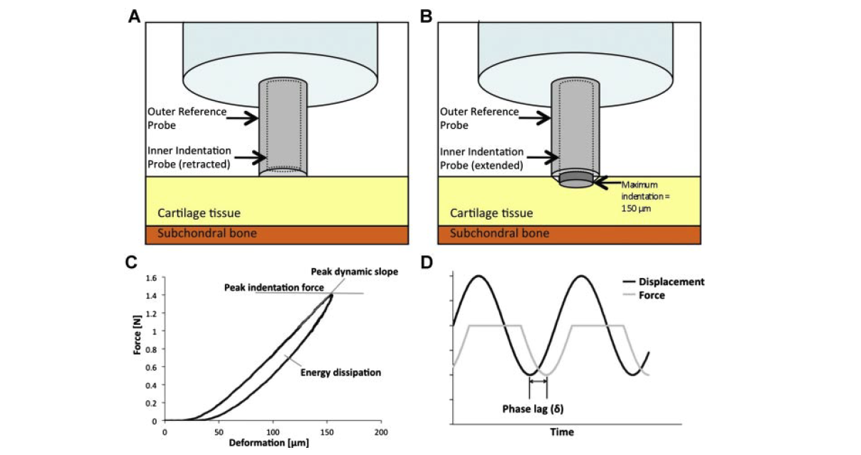
The objective of this study is to examine the local relationship between T(1ρ) relaxation times and the mechanical behavior of human osteoarthritic articular cartilage using high-resolution magnetic resonance imaging (MRI) and local in situ microindentation. Seven human tibial plateaus were obtained from patients who underwent total knee arthroplasty due to severe osteoarthritis (OA). Three to six sites were selected from each sample for visual classification using the ICRS Outerbridge scale (a total of 36 sites). Samples were imaged by MR, and the local distribution of T(1ρ) relaxation times were obtained at these selected sites. The elastic and viscoelastic characteristics of the tissue were quantified nondestructively using dynamic microindentation to measure peak dynamic modulus, energy dissipation, and phase angle. Measured Outerbridge scores, MR T(1ρ) relaxation times, and mechanical properties were highly heterogeneous across each cartilage surface. Site-specific measures of T(1ρ) relaxation times correlated significantly with the phase angle (p < 0.001; R = 0.908), a viscoelastic mechanical behavior of the cartilage. The novel combination of high-resolution MR imaging and microindentation allows the investigation of the local relationship between quantitative MRI and biomechanical properties in highly heterogeneous OA cartilage. These findings suggest that MRI T(1ρ) can provide a functional assessment of articular cartilage.
https://www.ncbi.nlm.nih.gov/pubmed/21445940
J Orthop Res. 2011 Sep;29(9):1312-9. doi: 10.1002/jor.21381. Epub 2011 Mar 28.
