
Abstract
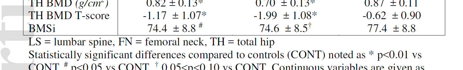
We tested whether cortical bone tissue properties assessed by in vivo impact microindentation would distinguish postmenopausal women with recent distal radius (DRF) or hip fracture (HF) from nonfracture controls (CONT). We enrolled postmenopausal women with recent DRF (n = 57), HF (n = 41), or CONT (n = 93), and used impact microindentation to assess bone material strength index (BMSi) at the anterior surface of the mid-tibia diaphysis. Areal bone mineral density (aBMD) (g/cm2 ) of the femoral neck (FN), total hip (TH), and lumbar spine (LS) were measured by dual-energy X-ray absorptiometry (DXA). HF and DRF subjects had significantly lower BMD than CONT at all sites (-5.6% to -8.2%, p < 0.001 for all). BMSi was 4% lower in DRF compared to CONT (74.36 ± 8.77 versus 77.41 ± 8.79, p = 0.04). BMSi was similarly lower in HF versus CONT, but the difference did not reach statistical significance (74.62 ± 8.47 versus 77.41 ± 8.79, p = 0.09). Lower BMSi was associated with increased risk of DRF (unadjusted OR, 1.43; 95% CI, 1.02 to 2.00, per SD decrease, p = 0.04), and remained statistically significant after adjustment for age, age and BMI, and age, BMI, and FN BMD (OR = 1.48 to 1.55). Lower BMSi tended to be associated with HF, but only reached borderline significance (unadjusted OR = 1.39; 95% CI, 0.96 to 2.01, p = 0.08). These results provide strong rationale for future investigations aimed at assessing whether BMSi can predict fracture in prospective studies and improve identification of women at risk for fragility fractures.
https://www.ncbi.nlm.nih.gov/pubmed/29115684
J Bone Miner Res. 2018 Apr;33(4):621-626. doi: 10.1002/jbmr.3338. Epub 2017 Dec 5.
