
Abstract
BACKGROUND:
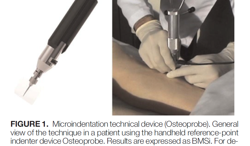
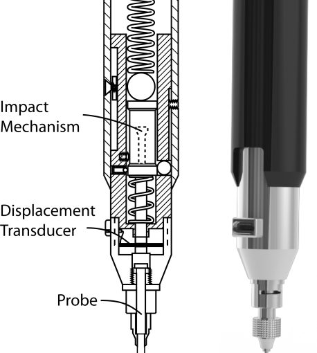
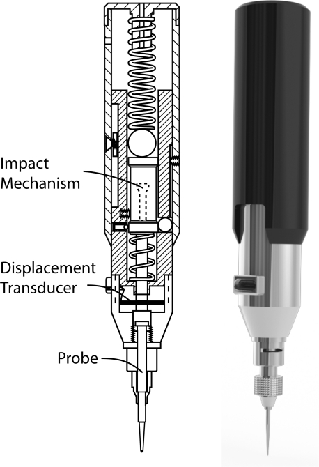
Bone mineral density (BMD) measured by dual-energy x-ray absorptiometry is used to assess bone health in kidney transplant recipients (KTR). Trabecular bone score and in vivo microindentation are novel techniques that directly measure trabecular microarchitecture and mechanical properties of bone at a tissue level and independently predict fracture risk. We tested the bone status of long-term KTR using all 3 techniques.
METHODS:
Cross-sectional study including 40 KTR with more than 10 years of follow-up and 94 healthy nontransplanted subjects as controls. Bone mineral density was measured at lumbar spine and the hip. Trabecular bone score was measured by specific software on the dual-energy x-ray absorptiometry scans of lumbar spine in 39 KTR and 77 controls. Microindentation was performed at the anterior tibial face with a reference-point indenter device. Bone measurements were standardized as percentage of a reference value, expressed as bone material strength index (BMSi) units. Multivariable (age, sex, and body mass index-adjusted) linear regression models were fitted to study the association between KTR and BMD/BMSi/trabecular bone score.
RESULTS:
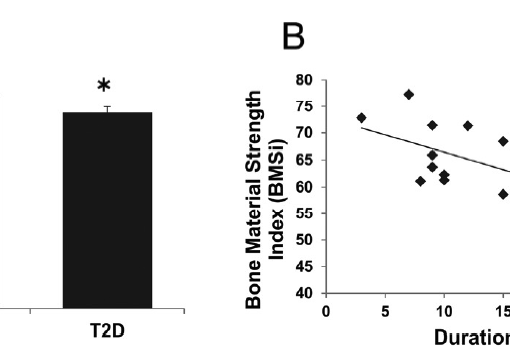
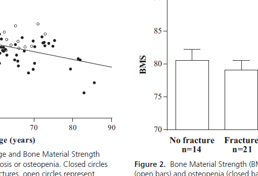
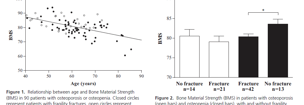
Bone mineral density was lower at lumbar spine (0.925 ± 0.15 vs 0.982 ± 0.14; P = 0.025), total hip (0.792 ± 0.14 vs 0.902 ± 0.13; P < 0.001), and femoral neck (0.667 ± 0.13 vs 0.775 ± 0.12; P < 0.001) in KTR than in controls. BMSi was also lower in KTR (79.1 ± 7.7 vs 82.9 ± 7.8; P = 0.012) although this difference disappeared after adjusted model (P = 0.145). Trabecular bone score was borderline lower (1.21 ± 0.14 vs 1.3 ± 0.15; adjusted P = 0.072) in KTR.
CONCLUSIONS:
Despite persistent decrease in BMD, trabecular microarchitecture and tissue quality remain normal in long-term KTR, suggesting important recovery of bone health.
https://www.ncbi.nlm.nih.gov/pubmed/27467533
Transplantation. 2017 Jun;101(6):1290-1294. doi: 10.1097/TP.0000000000001328.
