
Abstract
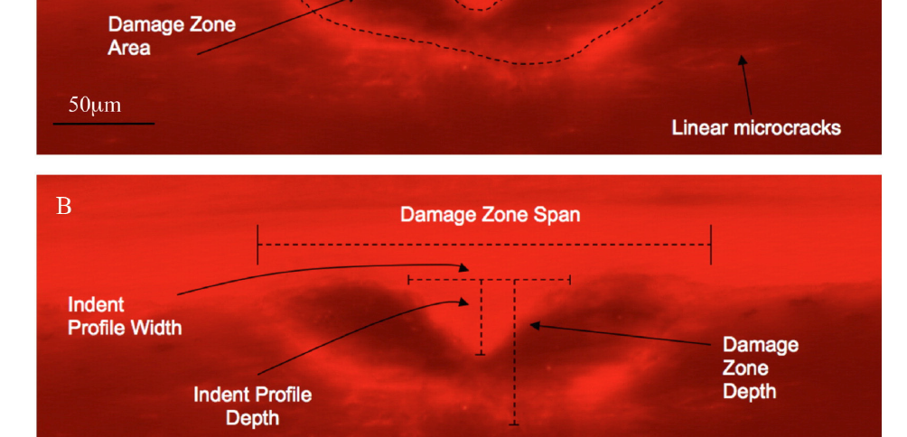
Measurement of bone mineral density (BMD) is the clinical gold standard in cases of compromised skeletal integrity, such as with osteoporosis. While BMD is a useful measurement to index skeletal health, it is also limited since it cannot directly assess any mechanical properties. The ability to directly assess mechanical properties of bone tissue would be clinically important. Reference point indentation (RPI) is a technology that has been designed to try and achieve this goal. While RPI has been shown to detect altered bone tissue properties, the underlying physical mechanism of these measurements has not been characterized. Thus, we designed a study whereby the contribution of (1) test cycle number and (2) test load level to RPI test-induced sub-surface damage was characterized and quantified. Standardized specimens were prepared from cadaveric human tibiae (n = 6), such that 12 replicates of each testing condition could be carried out. A custom rig was fabricated to accurately position and map indentation sites. One set of tests was carried out with 1, 5, 10, 15 and 20 cycles (Max Load: 8 N, Freq: 2 Hz), and a second set of tests was carried out with Load levels of 2, 4, 6, 8 or 10 N (Cycle number: 20, Freq: 2 Hz). The RPI parameter Loading Slope (LS) was cycle dependent at 5, 10, 15 and 20 cycles (p < 0.05). First Cycle Indentation Distance (ID 1st), Total Indentation Distance (TID), Mean Energy Dissipation (ED), First Cycle Unloading Slope (US 1st), Mean Unloading Slope (US) and LS were significantly different at 6, 8 and 10 N compared to 2 N (p < 0.05). From the histomorphometric measurements, damage zone span was significantly different after 5, 10, 15 and 20 cycles compared with 1 cycle while indent profile width and indent profile depth were significantly different at 10, 15 and 20 cycles (p < 0.05). With the load varying protocol, each of these parameters differed significantly at each increased load level (4, 6, 8, 10 N) compared with the basal level of 2 N (p < 0.05). The damage area parameter in both protocols was significantly different from baseline at the three upper levels tested (i.e. 10, 15, 20 cycles and 6, 8, 10 N, in cycle and load variant protocols, respectively). Specimens were scanned by micro-computed tomography, which showed no material or microstructural differences between samples, and processed for histological analyses and damage quantification. Consistent microdamage patterns were present with evidence of damage via compaction at the indented regions. While damage in the direction of loading was established early, the damage area then increased radially with cycle number. These data help to further understand the physical manifestations of RPI parameters and will help to further facilitate its use as a clinical diagnostic tool.
https://www.sciencedirect.com/science/article/pii/S8756328215000332
Bone Volume 75, June 2015, Pages 1-7 https://doi.org/10.1016/j.bone.2015.01.019